


The hip joint is the largest joint in our body.It has a hinge configuration that allows it to move in different planes.At the same time, the joint is surrounded by strong ligaments and muscles.The hip joints bear the main load when walking, running or carrying heavy loads.Coxarthrosis (another name for arthrosis of the hip joint) is quite common in people, both old and young.Once started, it may remain undiagnosed for a long time because visible limitation of hip movements does not occur immediately.
Often, patients, without being examined by a doctor or without presenting all their complaints, begin to treat lumbosacral osteochondrosis or arthrosis of the knee joints without any visible effect.Meanwhile, the untreated disease progresses and causes lameness, constant pain, shortening of the leg, and inability to bend and extend.And treatment at this stage is only possible surgically, that is, the joint has to be replaced with prostheses.
Causes of Coxarthrosis
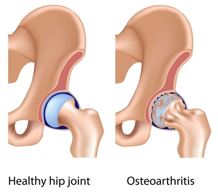
Primary arthrosis of the hip joint most often develops in people over 40 years of age.Its causes have not yet been studied.The hyaline cartilage that covers the joint surfaces and provides glide begins to thin and collapse.Due to increased friction and pressure on the bones, bone spurs appear.The joint is deformed, movements are limited.In primary coxarthrosis, both the knee joints and the spine are often affected.
Secondary arthrosis develops against the background of several diseases:
- Hip dysplasia.This term refers to the congenital underdevelopment of the components of this joint in a child.As a result, the femoral head is not centered as it should be in the acetabulum.There are three types of dysplasia: predislocation, subluxation, and hip dislocation.In congenital dislocation, the femoral head is located outside the socket and, if adequate treatment is not carried out, osteoarthritis subsequently develops.
- Aseptic necrosis.The bone tissue of the femoral head begins to dissolve due to impaired blood supply.Bone tissue is focally resorbed, the head of the joint is deformed.Arthrosis develops secondarily.
- Legg-Calvé-Perthes disease.This is osteochondropathy of the femoral head, which occurs in children aged 3 to 14 years, mainly in boys.It occurs, as a rule, as a result of complications following infectious processes, as well as injuries, physical overload and metabolic diseases.The cartilaginous region of the head is not well supplied with blood, which leads to necrosis of this area and deformation of the joint.
- Inflammation, infections.If arthritis of the hip joint develops, the synovial fluid loses its lubricating properties, the lining of the joint becomes thicker, the hyaline cartilage is subjected to mechanical stress, and at the same time metabolic disorders occur in the joint.
- Injuries: bruises, fractures of the femur, acetabulum, hip dislocations, chronic traumas, that is, microtraumas received systematically.
- Overload of the hip joint associated with sports and professional activities.For example, long walks without rest, vibration effects, constant jumping and carrying heavy loads are undesirable for the joint.The muscular corset of a child or teenager is not always able to compensate for such loads.
- Increase in body weight, especially at a young age, when the cartilage is not yet able to withstand large axial loads.Furthermore, these patients often have metabolic problems.
- Coxarthrosis itself is not inherited, but genetically relatives can have a certain structure of cartilaginous tissue, metabolic disorders that lead to the development of arthrosis.Therefore, it is worth considering whether parents or more distant relatives have joint diseases.
- Osteoporosis.The vulnerable area for this disease is the femoral neck.Its structure becomes more rarefied, making pathological fractures possible.All this secondary leads to osteoarthritis.
- Diabetes mellitus.In this case, arthrosis develops due to vascular disorders.
- Polyneuropathy with decreased sensitivity in the legs.
- Diseases of other parts of the musculoskeletal system.These include: scoliosis, osteoarthritis and knee injuries, flat feet.The distribution of load on the hip joints changes, shock-absorbing properties decrease and, as a result, the cartilaginous lining suffers.
Coxarthrosis symptoms
To prevent the disease and its early diagnosis, it is important to know the signs of incipient arthrosis of the hip joint (stage 1 coxarthrosis):
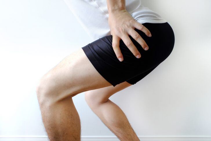
- Pain that occurs periodically after physical activity.Specific pain sensations may be localized to the groin, side, hip, or knee area.After rest they leave, so they are not given any importance.However, this is an alarming sign.
- Slight limitation of hip rotation (inward and outward).This can be easily checked while lying on your back by rotating your entire leg clockwise and counterclockwise.
- An x-ray may reveal a slight irregular narrowing of the joint space.
In stage 2 arthrosis, the signs are more pronounced:
- Pain occurs in the projection of the joint, most often in the inguinal fold, and is also observed at rest.
- Restrictions appear not only when rotating the leg, but also when abducting the hip to the side.Movements in the joint are somewhat painful, especially in extreme positions (with maximum abduction of the hip, flexion of the leg towards the stomach).
- On an x-ray, you can see a moderate narrowing of the joint space and isolated bony growths on the edges of the acetabulum.Cysts can also form in the bony structure of the femoral head.
Arthrosis of the hip joint of the 3rd stage is easily diagnosed, its symptoms are severe:
- Joint pain during exercise at night.
- Lameness, patients often use a cane.
- Marked limitation of joint movements, making it difficult for the person to put on socks or shoes.
- The leg becomes thinner due to hypotrophy of the thigh and lower leg muscles.The muscles in the gluteal region also weaken.
- It is possible to shorten the leg due to incomplete extension and deformation of the femoral head.As a result, scoliosis of the lumbar region (lateral curvature) forms and pain appears in the lumbosacral region.
- Signs of the 3rd stage, revealed by x-rays, are a pronounced narrowing of the joint space up to its complete absence, bone growths, deformation of the head and neck of the hip joint.
Diagnosis
In diagnosis, it is of great importance to clarify subjective complaints, collect anamnesis, evaluate symptoms and also clarify the stage - radiography, computed tomography and magnetic resonance imaging.Computed tomography allows you to study the bone structure of the hip joint in detail, and the magnetic resonance method visualizes the soft tissues, the state of the joint capsule and the presence of synovitis.
Treatment
Coxarthrosis therapy depends on the stage of the process and in most cases includes a whole range of procedures.It is clear that the sooner treatment is started, the greater its effectiveness.
- Conservative treatment
- Drug therapy.To relieve pain, non-steroidal anti-inflammatory drugs are used in tablets, suppositories or intramuscular injections.Pharmaceutical forms such as ointments, gels and creams are not effective enough due to the involvement of the hip joint by large muscles and subcutaneous tissue.Long courses of nonsteroidal anti-inflammatory drugs are not recommended due to side effects on the cardiovascular system and gastrointestinal tract.To help them, the doctor may prescribe medication that relieves muscle spasm - a muscle relaxant.In cases of severe inflammation, intra-articular glucocorticoids may be necessary.Chondroprotectors are one of the main groups of medications for treating coxarthrosis.They are administered intramuscularly and intraarticularly;In milder cases, tablets can be taken.These drugs are aimed at improving the restoration processes of cartilaginous tissue and slowing down its degeneration.The doctor may also prescribe vascular medications to improve local blood circulation.
- Physiotherapy.Their procedures improve blood flow in the joint area and relax muscles.These are UHF, magnetic therapy, laser treatment, diadynamic currents, electrophoresis.Purpose - according to individual indications.
- Therapeutic massage.An indispensable method of treating coxarthrosis: it relieves muscle spasms, has a beneficial effect on blood circulation and, when carried out systematically, strengthens the muscles.
- Therapeutic gymnastics.Improves blood flow and strengthens the muscular corset of the joint.Recommended exercises for coxarthrosis (performed on solid support):
- “bicycle” in supine position;
- lying on your back, hold your knee with your hand and pull it towards your stomach, and do the same with the other leg;
- lying on your back, bend your knees, press the soles of your feet on the floor and lift your pelvis, stay in this position;
- lying on your back, move your thigh to the side as much as possible;
- sitting on a chair, squeeze the ball between your thighs;
- lying on your back, turn your legs in and out;
- Standing with your right foot slightly elevated and holding the support with your hands, swing your left leg back and forth and left and right, then do the same, switching legs.
- Surgical treatment.Endoprosthesis, that is, the replacement of a joint with an artificial one, is performed in the 3rd stage of coxarthrosis in the presence of shortening of the limb, constant pain and severe contracture.Endoprostheses can be cemented (in the presence of osteoporosis) or cementless.The prosthesis itself can be unipolar (replacement of the head only) and total (replacement of both components).Already the day after the operation, some elements of exercise therapy are carried out lying in bed, the patient can stand, but for now without supporting the leg, and a few days later - with crutches.After 2-3 months, crutches will no longer be necessary and full weight bearing on the leg will be allowed.It is recommended that patients who have undergone endoprosthetics undergo rehabilitation consisting of physiotherapy, massage and physiotherapy.In most cases, limb function is restored.The service life of the prosthesis is 10 to 20 years, then it is replaced with a new one.
Coxarthrosis prevention
Prevention measures are very important, especially if you have a history of hip dysplasia, fractures, severe bruises or purulent processes in this area.
- Avoid lifting weights and jumping (especially from heights).Try not to stand for long periods of time.
- Controlling body weight (reduce the consumption of flour products, table salt, sweets, strong tea and coffee in the diet).Excess weight increases the risk of hip osteoarthritis.
- Dosed physical exercises aimed at strengthening the muscles of the thighs and glutes (cycling or stationary bike, swimming, therapeutic exercises).
- If there are diseases associated with metabolism (diabetes mellitus, atherosclerosis), they must be compensated.
Compliance with preventive measures, early detection of coxarthrosis and its appropriate treatment are the key to a positive prognosis for this disease.
Which doctor should I contact?
If you experience pain in your leg or hip joint, consult a doctor.He will prescribe primary diagnostic measures, in particular x-rays of the hip joint.Once the stage of the disease has been established, the patient will be referred to a rheumatologist or orthopedist.A nutritionist and endocrinologist can provide additional assistance in losing weight and slowing the progression of the disease.It would be helpful for women to consult a gynecologist to prescribe hormone replacement therapy to prevent osteoporosis.